Canadian Macromolecular Crystallography Facility - Bending Magnet
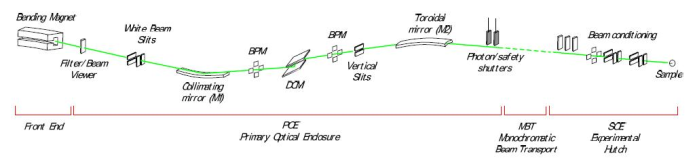
Designed to complement CMCF-ID, the CMCF-BM (08B1) bending magnet beamline is used for routine crystallographic analyses as well as more specialized techniques. The design incorporates white beam slits (WBS), a vertical collimating mirror (VCM), a double-crystal monochromator (DCM) / double-multilayer monochromator (DMM), and toroidal focusing mirror. The endstation includes a versatile Arinax MD2 microdiffractometer and Pilatus3 S 6M X-ray detector. A SAM automounter allows for remote operation, and data collection is controlled through MxDC.
The beamline is normally equipped with a Bruker XFlash 410 fluorescence detector to perform X-ray spectroscopy for MAD/SAD experiments and metal identification. A Vortex ME4 detector is alternatively available to perform specialized X-ray Absorption Near Edge Structure (XANES) and Extended X-ray Absorption Fine Structure (EXAFS) analyses on crystals. An HC1 humidity control device is available for use on the beamline, in place of the standard cryojet.
Modes
High Flux (DMM) Mode. The recent incorporation of the double-multilayer monochromator (DMM) allows high flux beam with energy adjustable between 7 - 10.5 keV (maximum flux @ 8.157 keV). This is the mode most commonly used for routine screening and data collection of native data. More information about this recent upgrade is available here.
Normal Flux (DCM or Si(111)) Mode. This lower flux mode requires longer exposure times, but is tuneable through the whole spectral range of the beamline (about 6 - 18 keV). This mode is most commonly used for MAD/SAD experiments or X-ray absorption spectroscopy experiments requiring energies outside the range of the high flux mode. It is also used when energies toward the upper end of the spectrum are needed for obtaining very high resolution data (very well-diffracting samples, small molecule crystallography and powder diffraction).
Note: Switching between the two modes is performed by staff (a process which normally takes about 5 - 10 minutes). Please confirm with your local contact as to which mode will be most appropriate for your work, or to change modes.
| Spectral Range (keV) |
DCM: 6 - 18 DMM: 7 - 10.5 (max flux @ 8.157) |
|---|---|
| Flux on Sample (ph/s) |
DCM: up to 1.5 x 10¹¹ @ 12 keV DMM: up to 2.5 x 10¹² @ 8.157 keV |
| Energy Bandwidth (ΔE/E) |
DCM: ~1.5 x 10⁻⁴ DMM: ~3.7 x 10⁻³ |
| Spot Sizes (µm) | 20, 50, 100, 150, 200 |
| Typical Ring Current (mA) | 220 (Top Up Mode) |
| Component | Distance from Source (m) |
|---|---|
| WBS (White Beam Slits) | 11.5 |
| Mirror M1 (Vertical Collimating) | 13.0 |
| DCM/DMM (Double Crystal/Double Multilayer Monochromator) | 17.1 |
| Mirror M2 (Toroidal) | 19.1 |
| Sample | 28.6 |
Hardware Details
| Bend Radius (m) | 7.14287 |
|---|---|
| Horizontal Fan Available (mrad) | 6 |
| Mirrors |
Collimating Mirror with two stripes (Si, Rh/Pt); Toroidal Focusing Mirror (Rh/Pt) |
|---|---|
| Horizontal Demagnification (1/Mx) | 2.01 (19.1:9.5) |
| Vertical Demagnification (1/My) | 1.37 (13:9.5) |
| Monochromator |
DCM: KOHZU Si₁₁₁ double crystal monochromator, featuring indirectly water-cooled first crystal and flat, long second crystal; DMM: Double multilayer monochromator featuring silicon substrates arranged in series with the DCM Si₁₁₁ crystals, Ni₉₃V₇/B₄C coating with 500 bilayers |
| Standard Pin Length (mm) | 18 |
|---|---|
| Cryo Capability | Oxford Instruments Cryojet5 (85 - 500K) |
| Automounter |
Stanford Automated Mounting system (SAM) with Unipucks, SSRL cassettes; Typical duty cycle < 30 s |
| X-Ray Fluorescence Detector | Bruker Xflash 410 |
| Additional Instrumentation |
Vortex ME4 X-Ray Fluorescence Detector; HC1 Humidity Control Device |
| Model |
Arinax MD2 Microdiffractometer; Optional MiniKappa |
|---|---|
| Maximum Rotation Speed (Deg/s) | 120 |
| Positioning Error (mDeg) | 2 |
| Sphere of Confusion (µm) | 2 |
| Type | On-Axis Visualization (OAV) |
|---|---|
| Orientation | Along beam axis |
| Maximum Zoom | 12x |
| Maximum Field of View (mm) | 1.8 x 1.5 |
| Minimum Field of View (mm) | 0.18 x 0.12 |
| Crystal-Detector Distance (mm) | 115 - 700 |
|---|---|
| Current Detector Model | Pilatus3 S 6M |
| Output file format | CBF |
| Framing Rate (Hz) | 25 |
| X-ray Sensitive Surface (mm) | 423.6 x 434.6 (184,097 mm²) |
| Head Dimensions (H x W x D) | 59.0 x 60.3 x 45.5 cm |
| 2Θ Range | 20° - not currently in use |
| Resolution | 2463 x 2527 |
| Detector History |
June 2020 - Present: Pilatus3 S 6M; August 25, 2015 - May 2020: Rayonix MX300HE; July 22, 2015 - August 20, 2015: Rayonix MX225; Before July 22, 2015: Rayonix MX300HE |